Pages

Tuesday, October 2, 2018

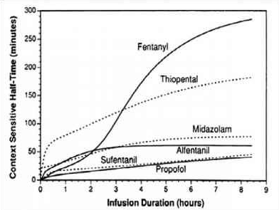
CONTEXT SENSITIVE HALF TIME [CSHT]

Screen Shot 2018-09-21 at 11.24.29 AM
  • Context sensitive half-time is deined as the time for the plasma concentration to fall to half of the value at the time of stopping an infusion
  • The half time will usually alter in the setting of varying durations  of drug infusion
  • The higher the ratio of distribution clearance to clearance due to elimination, the greater the range for context-sensitive half-time
  • The longest possible context-sensitive half-time is seen when the infusion has reached steady state, when there is no transfer between compartments and input rate is the same as elimination rate
  •  Draw and label the axes; draw the curve for the drug with the shortest CSHT first before plotting the others
  • REMIFENTANIL: Here the elimination always dominates distribution and so there is very little variation in CSHT with time and so it is context insensitive. Draw a straight line starting from the origin and becoming near horizontal after the CSHT reaches 5 min. This demonstrates that the half time is not dependent on the length of infusion as clearance by plasma esterases is so rapid. For remifentanil the
    longest possible CSHT is only 8 minutes
  • PROPOFOL: For propofol the clearance due to elimination is similar to that for distributioninto the second compartment, so plasma concentration falls rapidly after a propofol infusion mainly due to rapid eliminationwith a smaller contribution from distribution. Propofol is not context insensitive as its CSHT continues to rise; however it remains short even after prolonged infusions. Starting at the origin, draw a smooth curve rising steadily towards a CSHT of around 40 min after 8 h of infusion.
  • ALFENTANIL: The curve rises from the origin until reaching a CSHT of 50 min
    at around 2 h of infusion. Thereafter the curve becomes horizontal. This shows that alfentanil is also context insensitivefor infusion durations of 2 h or longer
  • THIOPENTONE SODIUM: The curve begins at the origin but rises more steeply than the others so that the CSHT is 50 min after only 30 min infusion duration. The
    curve should be drawn like a slightly slurred build-up exponential reaching a CSHT of 150 min after 8 h of infusion. As the CSHT continues to rise, thiopental does not become context insensitive
  • FENTANYLThe most complex curve begins at the origin and is sigmoid in shape. It should cross the alfentanil line at 2 h duration and rise to a CSHT of 250 min after 6 h of infusion. Again, as the CSHT continues to rise, fentanyl does not become context insensitive.
  • The maximum possible CSHT for propofol is about 20 minutes, compared with 300 minutes for fentanyl
  • It is important to realize that the CSHT does not predict the time to patient awakening but simply the time until the plasma concentration of a drug has fallen by half. The patient may need the plasma concentration to fall by 75% in order to awaken, and the time taken for this or any other percentage fall to occur is known as a decrement time.
  • Decrement time: The time taken for the plasma concentration of a drug to fall to the specified percentage of its former value after the cessation of an infusion designed to maintain a steady plasma concentration (time). The CSHT is, therefore, a form of decrement time when the specified percentage’ is 50%.
  • Although the CSHT for propofol has a maximum value of about 20 minutes, during long, stimulating surgery infusion rates will have been high and the plasma concentration when wake-up is required may be very much less than half the plasma concentration at the end of the infusion. Thus time to awakening using propofol alone may be much longer than the CSHT. This is why the TCI pumps display a decrement time rather than a CSHT.
  • When using propofol infusions, the decrement time is commonly quoted as the time taken to reach a plasma level of 1.2 μg.ml1, as this is the level at which wake up is thought likely to occur in the absence of any other sedative agents.
  • It must be remembered that after one CSHT, the next period of time required for plasma concentration to halve again is likely to be much longer. This relects the increasing importance of the slower redistribution and metabolism phases that predominate after re-distribution has taken place. This explains the emphasis on half-time rather than halflife: half-lives are constant whereas half-times are not!
MEDIASTENAL TUMOURS & THE ANESTHESIOLOGIST: SPECIFIC POINTS

  • A preoperative CT scan will show the site, severity, and extent of the airway compromise to assess the level and degree of obstruction.
  • Assess the vocal cord function preoperatively
  • Lung function tests to look for the extent of intrathoracic or extrathoracic obstruction.
  • ECHO to rule out pericardial effusion and cardiac compression.
  • Premedication with benzodiazepine is generally avoided if there is risk of airway compromise.
  • Airway equipment—rigid bronchoscopy and difficult airway trolley, jet ventilation, cardiopulmonary bypass (CPB) should be there as standby. Femoro femoral bypass is the most common setup.
  •  
  • COMPLICATIONS DUE TO MASS EFFECT OF THE TUMOUR: 
  •  
  • Vascular compromise—SVC Obstruction ( SVCO ) and pulmonary vessel obstruction
  • Laryngeal nerve palsy
  • Dysphagia
  • STRIDOR and airway compromise may be an important symptom
  • Inspiratory stridor (laryngeal)—obstruction above the level of glottis
  • Expiratory stridor (tracheobronchial)—obstruction in the intrathoracic airways
  • Biphasic stridor—obstruction between glottis and subglottis or a critical obstruction at any level
  • Sometimes you may have to go for a microlaryngoscopy tube (MLT)
  •  
  • TAKE CARE:
  • Aim to avoid worsening of cardiac compression, airway occlusion, and SVC obstruction.
  • IV cannula in the lower extremity
  • Induction in sitting position (semi Fowler’s position)
  • Inhalational (preferred choice) or IV induction agent titrated to effect
  • Choose spontaneous ventilation with LMA
  • Awake fibreoptic technique if intubation is necessary with a reinforced smaller calibre and longer endotracheal tube
  • Postoperative airway obstruction due to airway oedema, tracheomalacia, and bleeding warrant the need for awake extubation in ITU. The following steps would aid in an uneventful extubation:
  • Test for leak around the endotracheal tube cuff.
  • Administer dexamethasone or chemo radiotherapy in sensitive tumours to shrink size of tumour.
  • Use adrenaline nebulisers.
  • Extubate over airway exchange catheters.
  •  
  • SVCO: challenges during anaesthesia
  • Need for supplemental oxygen
  • Orthopnoea—induction in the sitting-up position
  • IV cannula in the lower extremity
  • Airway oedema
  • Mucosal bleeding
  • Laryngeal nerve palsy
  • Haemodynamic instability due to decreased venous return
  •  
  • OTHER CONCERNS 
  • General anaesthesia, causes loss of intrinsic muscle tone, decreased lung volumes, and decreased transpleural pressure gradient
  • Positive pressure ventilation, can precipitate severe hypotension and also increases intrathoracic tracheal compression
  • Coughing, as it can cause complete airway obstruction by positive pleural pressure, increasing intrathoracic tracheal compression
  • Following gas induction, the patient stops breathing and if you are unable to ventilate her: Follow difficult or failed intubation guidelines. But cricoid puncture and emergency tracheostomy are futile if the level of airway obstruction is at the intrathoracic tracheobronchial tree: Try a change in position—lateral, sitting up, or prone—to decrease the mechanical effect of the tumour. Avoid positive pressure ventilation for fear of luminal closure. Low-frequency jet ventilation with Sander’s injector or high-frequency translaryngeal jet ventilation with Hunsaker’s catheter is one option. CPB bypass and ECMO to restore oxygenation when other measures fail.
  • Following chemotherapy in ICU, if patient develops hyperkalemia, Tumour Lysis Syndrome should be there in the differential diagnosis
  • ALSO NOTE
  • During inspiration, the intrathoracic airways expand along with the expanding lungs. In contrast, the extrathoracic airways diminish in caliber during inspiration due to their intraluminal pressure being lower than the atmospheric pressure. The reverse happens during expiration.
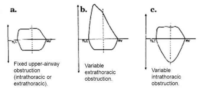
  • Flow volume loop inupper-airway obstruction:
  • Fixed lesions[extrathoracic or intrathoracic] are characterized by lack of changes in caliber during inhalation or exhalation and produce a constant degree of airflow limitation during the entire respiratory cycle. Its presence results in similar flattening of both the inspiratory and expiratory portions of the flow-volume loop
  • Variable lesions are characterized by changes in airway lesion caliber during breathing. Depending on their location (intrathoracic or extrathoracic), they tend to behave differently during inhalation and exhalation.
  • In the case of an extrathoracic obstructing lesion, during inspiration, there is acceleration of airflow from the atmosphere toward the lungs, and the intraluminal pressure decreases with respect to the atmospheric pressure due to a Bernoulli effect, resulting in the limitation of inspiratory flow seen as a flattening in the inspiratory limb of the flow-volume loop. During expiration, the air is forced out of the lungs through a narrowed (but potentially expandable) extrathoracic airway. Therefore, the maximal expiratory flow-volume curve is usually normal.
  • Variable intrathoracic constrictionsexpand during inspiration, causing an increase in airway lumen and resulting in a normal-appearing inspiratory limb of the flow-volume loop. During expiration, compression by increasing pleural pressures leads to a decrease in the size of the airway lumen at the site of intrathoracic obstruction, producing a flattening of the expiratory limb of the flow-volume loop
  • Screen Shot 2018-09-19 at 1.34.27 AM

ETOMIDATE: THE BAD 'WOW' FACTORS

  • While it continues to be used infrequently in the UK it has been withdrawn in North America and Australia
  • The most notable and potentially serious side effect of etomidate administration is the suppression of adrenocortical steroid synthesis.
  • It suppresses adrenocortical function by inhibition of the enzymes 11-hydroxylase and 17-hydroxylase, resulting in inhibition of cortisol and aldosterone synthesis.
  • After a single bolus dose of etomidate, this adrenocortical suppression lasts approximately 6 h in healthy individuals. However in the critically ill, such suppression can last for days. In other words the situation in which it has the best cardiovascular proile is the unwell patient in whom the consequences of steroid inhibition are likely to be the most detrimental.
  • Etomidate is approximately 100-fold more potent a suppressor of adrenocortical function than it is a sedative-hypnotic. Consequently, an anesthetic
    induction dose of etomidate represents a massive overdose with respect to its ability to suppress adrenocortical function. And etomidates terminal elimination half-life is rather long. Thus, after just a single anesthetic induction dose of etomidate, many hours must pass before etomidates concentration in the blood falls below that which suppresses adrenocortical steroid synthesis.
  • It is within this mechanistic context that the strategy emerged to design analogues of etomidate
  • ANALOGUES:
  • MOC-etomidate[ relatively low potency and very rapid metabolism (1.) required the administration of extremely large doses]
  • CPMM etomidate[ it has an onset and offset of hypnotic action that are fast (1.) ]
  • Carbo etomidate[ less adrenocortical inhibition (2.)]
  • MOC-carboetomidate[ combines properties (1) and (2); but it's potency
    is very low which means that extremely large doses would need to be administered to maintain anesthesia ]
  • Involuntary movements (myoclonus) are commonly observed after etomidate administration, with some studies reporting an incidence as high as 80 % in unpremedicated patients
  • It has been suggested that it occurs because etomidate depresses inhibitory neural circuits in the central nervous system sooner and at lower concentrations than excitatory circuits.
  • Regardless of the mechanism, myoclonus can be significantly reduced or completely prevented by administering a variety of drugs with central nervous system depressant effects including opiates, benzodiazepines, dexmedetomidine, thiopental, lidocaine, and magnesium.
  • Pain at the injection site is another common side effect and its incidence is highly dependent upon the size of the vein into which it is injected and the formulation that is used.
  • Lipid emulsion and cyclodextrin formulations may reduce TRP channel activation, leading to less pain on injection
  • Postoperative nausea and vomiting is common with reported incidences as high as 40 %. It has been suggested that the emetogenic trigger in etomidate is the propylene glycol solvent and not the anesthetic itself.
  •